Meet my Scottish Terrier friend Spondi!
We learned in Medical Imaging today what a useful tool he is in visualizing the lumbar spine in x-rays and picking up a particular fracture called a “spondylolysis” (phew, say that 5 times fast).

A spondylolysis is a defect of the pars interarticularis of the vertebral arch in lumbar vertebrae (most commonly L5 but it can happen more proximally too), so check out this x-ray and see if you can see my friend Spondi on this oblique view of the L-Spine:

ahhh, well a little dotted line never hurt. Look at his cute little nose, ears, collar, and tail.
So here are some times. Spondi faces the side of the x-ray (which is relative to the patient) so the dog in the x-ray above is facing to the patients left. The gots eyes are the pedicles, the collar is the par interarticularis and it will be more radiolucent with separation, and his nose the transverse process. Pretty cool huh?!
How about another drawing for clarification:
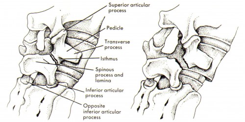
(image from http://www.athleticadvisor.com/injuries/head_back/back_anatomy.htm)
