If you know any graduate students (especially in the medical profession) we have A GAZZILION pneumonics to remember all the bones, muscles, pathologies, etc, etc … some how they are always a little, well, PG-13. SOOOOO, now that the title of this post has grabbed your attention, we will not be discussing bondage –> instead how about a little MRI interpretation? I know, I know…not as “loin quivering” but bear with me.
Very simply, MRI (magnetic resonance imaging) uses large, powerful magnets to align a magnetic field interacting with atomic nuclei in the body which is then interpreted by a computer to help clinicians visualize the slices of the body. They can often look similar to a CT scan, but remember that MRIs often show soft tissue anatomy a bit clearer while CTs are more ideal for looking at bones.
There are two common ways to “weight” MRIs
T-1 uses a short time between the pulse and signal capture. T-1 gives you the clearest picture of anatomy, with structures like fat & white matter showing up bright white (intense).
T-2 uses a longer time period to take the picture, so parts of the body such as water/fluid that are slow to interact with the pulse are clearer. This makes them GREAT to visualize inflammation.
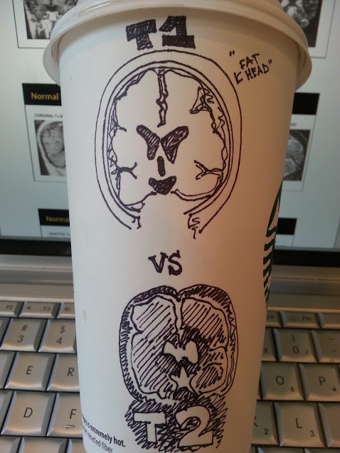
So check out this coronal slice of the brain.
Ventricles in T-1 are going to be DARK, while in T-2 they are WHITE (fluid)
Another give away, is the subcutaneous fat in the T-1 lining the skull.
So, quiz yourself. How are these MRIs weighted? Hover over ’em for the answers:


